
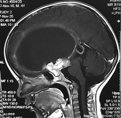
Giant Craniopharyngioma
Abstract
Background: Pediatric craniopharyngiomas are adamantinomatous craniopharyngioma. They have solid and cystic parts. They can achieve enormous sizes.
Clinical presentation: They present with headache, visual disturbances, cognitive decline, hormonal deficiency and short stature.
Key diagnostic features: They are heterogenous suprasellar solid masses with cystic components. The solid part enhances. Calcification is common. They cause sellar erosion.
Differential diagnosis:
Rathke cleft cyst-no solid or enhancing component, calcification is rare, unilocular, the majority are completely or mostly intrasellar.
Pituitary adenoma -usually has intrasellar epicenter with sellar enlargement rather than the suprasellar epicenter, calcification is often absent.
Teratoma - presence of fat is helpful but requires fat-saturated sequences or CT to confirm.
Hypothalamic/optic pathway gliomas-homogenous infiltrating mass which enhances on contrast.
Treatment: Surgical debulking with or without radiotherapy is the mainstay. Recurrence is seen in about 50% patients. Intracystic bleomycin is also useful.
Downloads
References
AJNR Am J Neuroradiol. 2020 Jan; 41(1): 106–110.
2. Lee HJ , Wu CC , Wu HM , Hung SC , Lirng JF , Luo CB , Chang FC , Guo WY . Pretreatment diagnosis of suprasellar papillary craniopharyngioma and germ cell tumors of adult patients. NR Am J Neuroradiol. 2015 Mar; 36(3):508-17

This work is licensed under a Creative Commons Attribution 4.0 International License.


